Williams Lab
We investigate how gene regulatory networks and signalling pathways govern lineage segregation at the Neural Plate Border — directing cells toward neural crest and cranial placode fates in the early embryo.
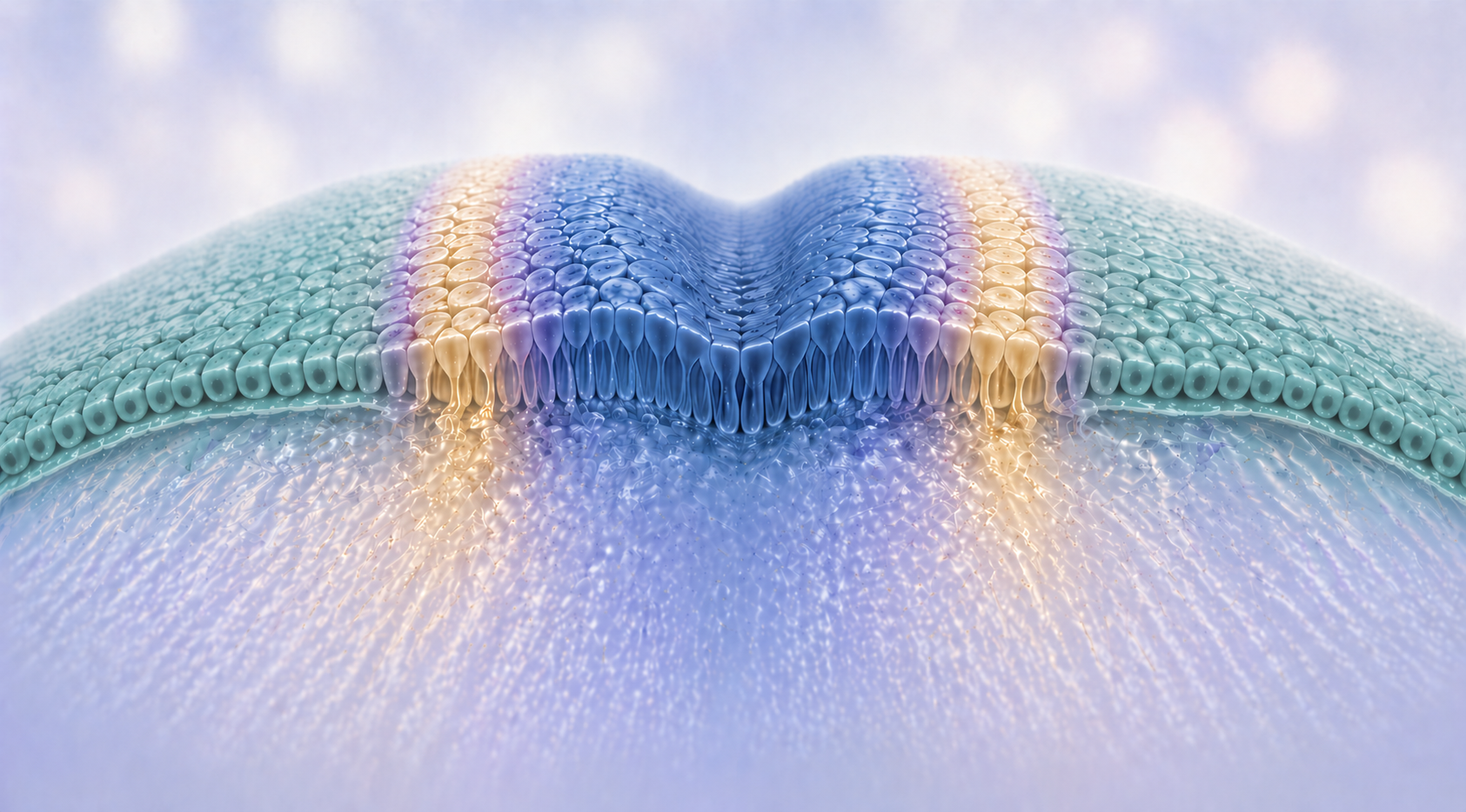
Neural crest cells are a remarkable, multipotent stem cell-like population that migrates extensively and gives rise to diverse derivatives including peripheral neurons, glia, pigment cells and craniofacial cartilage and bone. Cranial placodes are specialised ectodermal thickenings that produce the sensory organs of the head — the olfactory epithelium, lens, inner ear and cranial sensory ganglia — making them essential for vertebrate sensory function.
Defects in neural crest and placode development lead to syndromic birth defects, and embryonic gene regulatory networks are known to be reactivated in several cancers. Therefore, a thorough understanding of the molecular intricacies underlying the behaviour of these fascinating cells will help uncover the causes and potential therapeutics for such conditions.